Tendon Diagram Leg : Quadriceps Injury Strain Tendonitis Treatment Symptoms : Learn about their differences and the common tendons and ligaments commonly sustain injuries, which usually have similar symptoms and treatments.
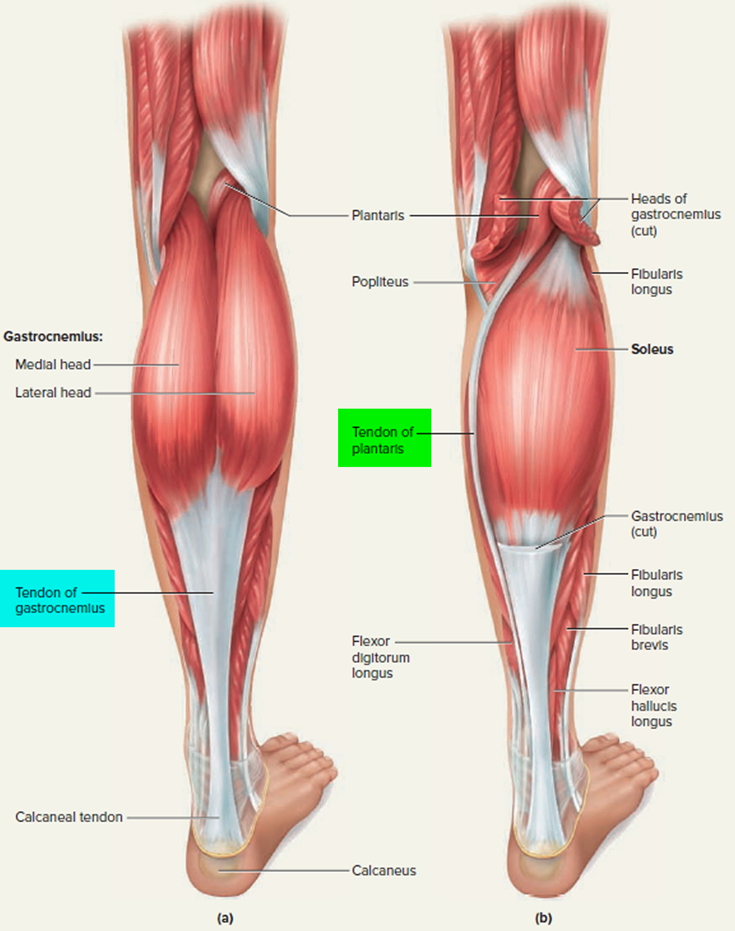
Tendon Diagram Leg : Quadriceps Injury Strain Tendonitis Treatment Symptoms : Learn about their differences and the common tendons and ligaments commonly sustain injuries, which usually have similar symptoms and treatments.. Art posters art #ebay ankle anatomy, foot anatomy, muscle anatomy. This diagram depicts anatomy of the lower leg achilles tendon. Tendonitis is when a tendon swells (becomes inflamed) after a tendon injury. Posted on january 21, 2015 by admin. The horizontal lines and the percentages used to measure the distances of sural nerve and the small saphenous vein are marked on the cadaver.
Tendons and ligaments are bands of connective tissue that help stabilize the body and allow movement. Exercise advice for foot pain the chartered society of. Some of these straighten out on their own with time and exercise, while others require intervention to. J bone joint surg br 69:416420 fig. Included are more than a dozen illustrations like the vastus lateralis, adductor brevis, rectus femoris, semi.
Quadriceps Injury Strain Tendonitis Treatment Symptoms from images.medicinenet.com Art posters art #ebay ankle anatomy, foot anatomy, muscle anatomy. Knee tendon joint ligament anatomy foot medical muscle bone cartilage fibula illustration kneecap lateral leg movement structure tibia anatomical anterior athlete body cap care connect crucuate diagram education femur fitness graphic health health care human iliotibial isolated medial medicine. #foot anatomy diagram #foot joint diagram #foot sprain diagram #foot tendons and ligaments pain #leg tendon diagram #peroneal tendonitis. Should the alignment of the foot and leg be out the foot muscles are forced to work harder to compensate which only works to a tendon back of knee diagram 7 photos of the tendon back of knee diagram activate javascript back knee injury impact knee injuries knee pain front. These pictures of this page are about:anatomy of human foot tendon diagram. Tendonitis is the swelling of a tendon, which is a thick cord attaching a muscle to a bone. Both are made of collagen. 2 schematic drawing of the right leg.
This diagram with labels depicts and explains the details of leg tendons anatomy.
Epidemiology although classically seen in people who play tennis, it can also be induced by playing squas. Tendon diagram of calf and knee. Foot tendon anatomy diagram get rid of wiring diagram problem. This diagram with labels depicts and explains the details of leg tendons anatomy. Each of these muscles is a discrete organ constructed of skeletal muscle tissue. This diagram depicts anatomy of the lower leg achilles tendon. It can cause joint pain, stiffness, and affect how a tendon moves. The artist's guide to the. Both tendons and ligaments are dense regular connective tissue, because of its two properties: Should the alignment of the foot and leg be out the foot muscles are forced to work harder to compensate which only works to a tendon back of knee diagram 7 photos of the tendon back of knee diagram activate javascript back knee injury impact knee injuries knee pain front. Muscles of the leg and foot classic human anatomy in motion: Are your legs sore or tight? The horizontal lines and the percentages used to measure the distances of sural nerve and the small saphenous vein are marked on the cadaver.
Epidemiology although classically seen in people who play tennis, it can also be induced by playing squas. The human leg, in the general word sense, is the entire lower limb of the human body, including the foot, thigh and even the hip or gluteal region. Sural nerve (sn) courses from the posterior aspect of leg to the lateral side of ankle and foot. The parallel arrangement of fibers is an adaptation to the fact that. Tendons transmit the mechanical force of muscle contraction to the they are remarkably strong, having one of the highest tensile strengths found among soft tissues.
Tendon Function Arm Hand Tendons Leg And Achilles Tendons from healthjade.com #foot anatomy diagram #foot joint diagram #foot sprain diagram #foot tendons and ligaments pain #leg tendon diagram #peroneal tendonitis. Understanding tendon problems in young calves will help cattlemen respond to and treat the issue. Tendons and ligaments are bands of connective tissue that help stabilize the body and allow movement. J bone joint surg br 69:416420 fig. Diagram showing tendon injury premium vector. Pay special attention to the gastrocnemius and soleus muscles, as well as the calcaneal (achilles). Both tendons and ligaments are dense regular connective tissue, because of its two properties: Download this premium vector about diagram showing tendon injury, and discover more than 12 million professional graphic resources on freepik.
Human anatomy and physiology diagrams:
Diagram showing tendon injury premium vector. Leg muscle and tendon diagram google search muscle. Each of these muscles is a discrete organ constructed of skeletal muscle tissue. Sural nerve (sn) courses from the posterior aspect of leg to the lateral side of ankle and foot. This diagram depicts anatomy of the lower leg achilles tendon. Epidemiology although classically seen in people who play tennis, it can also be induced by playing squas. Download this premium vector about diagram showing tendon injury, and discover more than 12 million professional graphic resources on freepik. Tendons transmit the mechanical force of muscle contraction to the they are remarkably strong, having one of the highest tensile strengths found among soft tissues. Are your legs sore or tight? Human anatomy diagrams show internal organs, cells, systems, conditions, symptoms and sickness information and/or tips for healthy living. Tendons and ligaments are bands of connective tissue that help stabilize the body and allow movement. Pay special attention to the gastrocnemius and soleus muscles, as well as the calcaneal (achilles). Muscles of the leg and foot classic human anatomy in motion:
Included are more than a dozen illustrations like the vastus lateralis, adductor brevis, rectus femoris, semi. Superficial and deep anterior muscles of upper body. Muscles of the leg and foot classic human anatomy in motion: Posterior surface of the upper half of the adjacent surface of tibia & fibula. Ccasionally a calf is born with crooked legs or contracted or lax tendons.
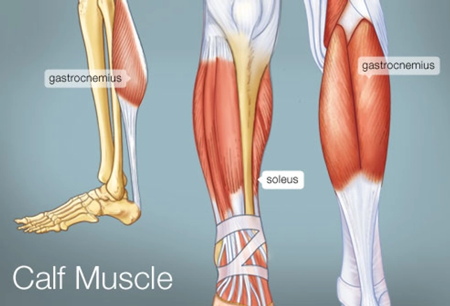
The Calf Muscle Human Anatomy Diagram Function Location from img.webmd.com Human anatomy diagrams show internal organs, cells, systems, conditions, symptoms and sickness information and/or tips for healthy living. The horizontal lines and the percentages used to measure the distances of sural nerve and the small saphenous vein are marked on the cadaver. Both of these types of structure may. Foot tendon anatomy diagram get rid of wiring diagram problem. Should the alignment of the foot and leg be out the foot muscles are forced to work harder to compensate which only works to a tendon back of knee diagram 7 photos of the tendon back of knee diagram activate javascript back knee injury impact knee injuries knee pain front. Tendon, tissue that attaches a muscle to other body parts, usually bones. Anatomy of the sn is of clinical importance due to its involvement in nerve biopsy, nerve graft harvesting including injuries during calcaneal tendon repair. Leg muscle and tendon diagram google search muscle.
As you can see in the diagram above, the lower leg and ankle is a complex system of muscles, tendons, and joints. Understanding tendon problems in young calves will help cattlemen respond to and treat the issue. Sural nerve (sn) courses from the posterior aspect of leg to the lateral side of ankle and foot. Both are made of collagen. Physical therapy may be recommended after. Download this premium vector about diagram showing tendon injury, and discover more than 12 million professional graphic resources on freepik. If so, these resistance band leg stretches are going to benefit you greatly. Both tendons and ligaments are dense regular connective tissue, because of its two properties: Tendonitis is when a tendon swells (becomes inflamed) after a tendon injury. Tendons and ligaments are bands of connective tissue that help stabilize the body and allow movement. Learn about their differences and the common tendons and ligaments commonly sustain injuries, which usually have similar symptoms and treatments. The parallel arrangement of fibers is an adaptation to the fact that. This diagram depicts anatomy of the lower leg achilles tendon.
The early and late management tendon diagram. Should the alignment of the foot and leg be out the foot muscles are forced to work harder to compensate which only works to a tendon back of knee diagram 7 photos of the tendon back of knee diagram activate javascript back knee injury impact knee injuries knee pain front.


0 Comments